Elbow Arthroscopy
Elbow Surgery examples
Patient is an active 55 year old male with persistent pain to the inside of his elbow and instability to his ulnar nerve
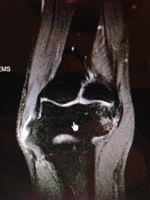
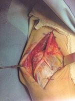
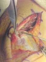
Patient is an active 55 year old male with persistent pain to the inside of his elbow and instability to his ulnar nerve. After failing conservative therapy the patient elected to undergo debridement and repair of his common flexor origin in addition to subcutaneous ulnar nerve transposition. Figure 1 is the patient's MRI which demonstrates the tearing at the medial epicondyle where the common flexor origin attaches to the humerus. Figure 2 demonstrates the hole or tearing in the common flexor origin at the time of surgery – this tear was debrided and then reattached to the humerus with the use of a suture anchor. Figure 3 shows the final surgical result with the ulnar nerve transposed.
Valgus Extension Overload in a Professional Pitcher
The patient is a 26 year-old professional baseball player with increasing pain with follow through. He presents with pain in the medial aspect of the posterior compartment. His exam is consistent with classic valgus extension overload. His MRI demonstrates loose pieces posteriorly along with a large posterior medial osteophyte (MRI1 and MRI2). Arthroscopic pictures reveal two large loose pieces in the posterior compartment of the elbow (AS1, AS2). The large posterior osteophyte was loose and the source of his pain with pitching (AS3, Phyte video). The loose fragments and osteophyte were removed (AS4). The tip of olecranon was resected as well to decrease pain with terminal extension / follow through. (AS5) The patient returned to pitching at 6 weeks post operation.
Phyte Video
Medial Epicondyle Avulsion Fracture
The patient is a 16 year old softball athlete who felt a pop while throwing to 1st base. She has avulsed her medial epicondyle via the pull of the mediall epicondyle. At surgery, the ulnar nerve was decompressed, but was not transposed. A suture is place around the common flexor origin to get control of the fracture fragment. A 4.5 mm screw is inserted into the medial epicondyle to hold it in its anatomic position while it heals. She returned to unrestricted softball in 3 months and played for another 4 years at the collegiate level without difficulty.
Click on the thumbnails for enlarged view
Arthroscopic Elbow Arthrofibrosis Release
Arthrofibrosis (stiff joint) is a common reason people come to see Dr Shepard. Using four to six poke holes in the skin and a combination of cameras (2.7 mm, 4.5mm), Dr Shepard will remove the loose bodies, bone spurs, and the contracted capsule (casing) that restrict the elbows range of motion. Less than 10% of Orthopaedic Surgeons perform elbow arthroscopy and even fewer perform arthroscopic releases. An arthroscopic release caries several advantages over traditional open releases: less pain, quicker recovery, better final range of motion.
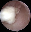
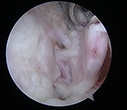
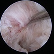
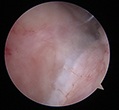
This patient presented with severe loss of motion - her range of motion was 30-95 degrees, whereas normal motion is 0 (straight) to 145 (bent) degrees. Picture 1 shows the scarred anterior capsule, which was restricting extension. Pictures 2, 3 and videos 1,2 show the anterior capsule being resected. Picture 4 shows the removal of a loose body that was causing irritation and pain. Pictures 5,6 and video 3 show resection of the olecranon tip / bone spurs and the resection of the posterior capsule. Final motion achieved with the release was 0-135.
Click on the thumbnails for enlarged view
Video 1
Video 2
Video 3
Subcutaneous Ulnar Nerve Transposition
Ulnar Nerve Transposition is performed for irritation of the ulnar nerve in the cubital tunnel and or recurrent subluxation of the nerve out of the tunnel. In this patient, the nerve was unstable during simple elbow range of motion and while playing water polo.
Video 1
An incision is made directly over the medial epicondyle - allowing access to the nerve proximally in the arm between the brachialis and the triceps, as well as access distally as the nerve enters the forearm. Care is taken during the dissection to isolate the medial antebrachial cutaneous nerve which runs across the incision. A wide dissection is neccessary to adequately decompress the nerve.
Video 2
The nerve is idenitifed proximal to the medial epicondyle and protected with a vessel loop. Distally, the nerve penetrates the two heads of the flexor carpi ulnaris muscle and this muscle is carefully split to allow decompression and transposition of the nerve.
Video 3
The ulnar nerve is transposed out of the cubital tunnel and lays atop the common flexor origin. A sling is fashioned from the fascia overlying the common flexor origin to gently prevent recurrent subluxation. Finally, the triceps fascia is reapproximated to the cubital tunnel to "close the tunnel" and ensure the nerve could not fall back into the tunnel.
How is elbow arthroscopy different than other surgeries?
Elbow arthroscopy is not as common a procedure as arthroscopy of the knee and shoulder. According to the American Academy of Orthopaedic Surgeons Survey, less than 10% of orthopaedic surgeons perform elbow arthroscopy. Elbow arthroscopy is technically demanding, requires multiple portals, utilizes multiple cameras, and should be performed with someone with a strong background in the surgery. Dr Shepard learned elbow arthroscopy from world famous James R Andrews MD – one of the developers and forefathers of the technique in the United States. Dr Shepard has performed elbow arthroscopy on high level overhead athletes from UC Irvine, Cal State Fullerton, and Chapman Universities.
Who needs elbow arthroscopy?
There are two primary groups of people who undergo elbow arthroscopy – young overhead athletes with loose bodies and OCD lesions; older people with loss of motion (arthrofibrosis).
The young athletes often develop loose bodies or cartilage lesions from their repetitive injuries. These lesions can be treated through the arthroscope with loose body removal and cartilage restoration techniques. These injuries are most commonly seen in baseball and softball athletes, as well as young athletes who compete in gymnastics, wrestling, and weight lifting.
The older athlete who looses motion, also known as arthrofibrosis, can be treated through the arthroscope as well. These patients loose the function of their elbow as their motion decreases over time. This problem can be treated with an arthroscopic release of scar tissue and capsule to restore motion. Sometimes, a small burr will be used to resect bone spurs to restore motion.


